


Pterygopalatine Fossa Tumour Management
by Premier Hospitals | January 22, 2024 |
Table Of Contents
- Introduction: Pterygopalatine Fossa Tumour Management
- Patient Presentation: Understanding Cases at Premier Hospital
- Diagnosis: Precision in Identifying Tumours
- Treatment: Tailored Approaches for Effective Management
- Surgical Steps: Precision Procedures in Tumour Removal
- Meet the Surgeon: Profiling the Expert Behind Successful Surgeries
- Conclusion: Achieving Success in Pterygopalatine Fossa Tumour Management
Introduction to Pterygopalatine Fossa Tumour Management at Premier Hospital
Pterygopalatine Fossa Tumour' refers to a tumour situated in a small space behind the upper jaw, known as the Pterygopalatine Fossa. This area contains important structures like nerves, blood vessels, and connective tissues. Tumours in this fossa can vary, ranging from benign to malignant, and may originate from different tissues, influencing symptoms, treatment options, and prognosis.
Due to the complex anatomy and delicate structures in the Pterygopalatine Fossa, surgical management often involves specialised techniques such as swing maxillotomy. This surgical approach improves access to the fossa by temporarily moving the upper jaw away from the skull, providing better visibility and flexibility during the procedure.
In this article, we present the management of an uncommon case—a left pterygopalatine fossa tumour in one of our patients. Our skilled medical team at Premier Hospital, Hyderabad, addressed this challenge adeptly, utilising the swing maxillotomy technique alongside gross total excision.
Patient Presentation in Pterygopalatine Fossa Tumour Management Cases
The patient, a 26-year-old female, presented with a persistent complaint of left cheek pain endured for a year. Over the last two months, the pain escalated, leading her to seek medical attention. Notably, she experienced tingling and numbness in the left cheek, adding complexity to her condition.
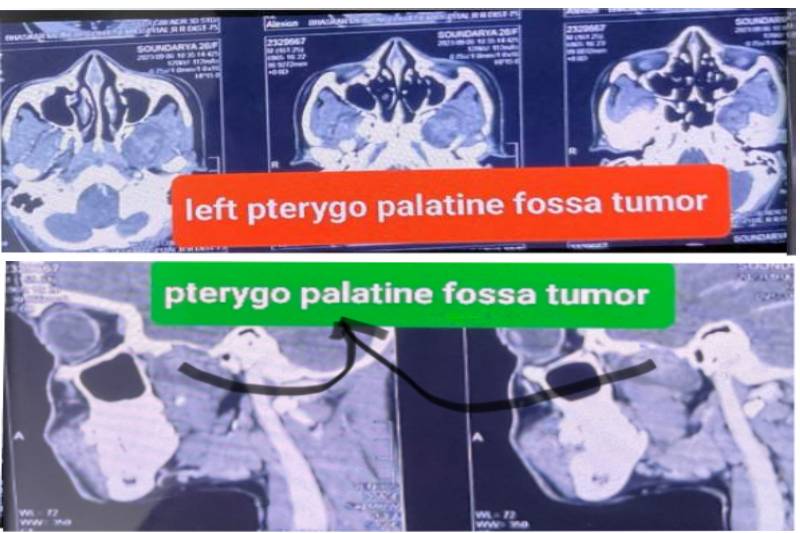
The team at Premier Hospital, led by a neurosurgeon, conducted a series of tests to comprehensively evaluate the nature of her symptoms. These tests included MRI of the brain and paranasal sinuses and CT imaging of the paranasal sinuses. These investigations played a crucial role in uncovering the details of the left pterygopalatine fossa tumour and guiding the subsequent course of management.
Diagnosis for Pterygopalatine Fossa Tumour Management: Precision in Identifying Tumours
Upon initial examination, the patient presented with good health, maintaining a fair general condition and displaying no fever or abnormalities in vital signs. Importantly, there were no apparent focal neurological deficits observed.
Further evaluation through MRI of the brain and paranasal sinuses, along with CT imaging of the paranasal sinuses, revealed compelling evidence. The scans depicted a well-enhancing lesion in the left pterygopalatine fossa, with its lateral margins extending toward the left Internal Carotid Artery (ICA). This radiological evidence suggested the possibility of a Schwannoma, potentially originating from the V2 branch of the left maxillary nerve.
The diagnostic findings unveiled by the imaging studies served as crucial pieces in unravelling the nature and extent of the left pterygopalatine fossa tumour.
Treatment Strategies in Pterygopalatine Fossa Tumour Management: Tailored Approaches for Effective Results
The patient, presenting with a year-long history of left cheek pain intensifying over the last two months, accompanied by tingling and numbness, underwent a diagnostic journey that culminated in revealing a well-enhancing lesion in the left pterygopalatine fossa, extending towards the left Internal Carotid Artery—suggestive of a potential Schwannoma arising from the V2 branch of the left maxillary nerve.
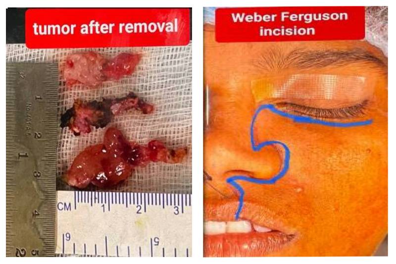
Subsequently, a Weber Ferguson incision paved the way for a left swing maxillotomy, enabling the meticulous gross total excision of the tumour on October 26, 2023. Postoperatively, the patient exhibited swift mobilisation the next day, initiating Ryle’s tube feeding and clear oral fluids.
Surgical Steps in Pterygopalatine Fossa Tumour Management: Precision Procedures in Tumour Removal
The surgical intervention for the left pterygopalatine fossa tumour comprised a series of meticulous steps, ensuring both precision and efficacy.
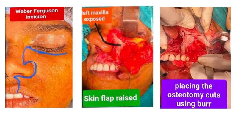
- Weber-Ferguson Incision: - A Weber-Ferguson incision was made on the left side, a commonly used technique in craniofacial surgery, providing optimal access to the oral and maxillofacial regions.
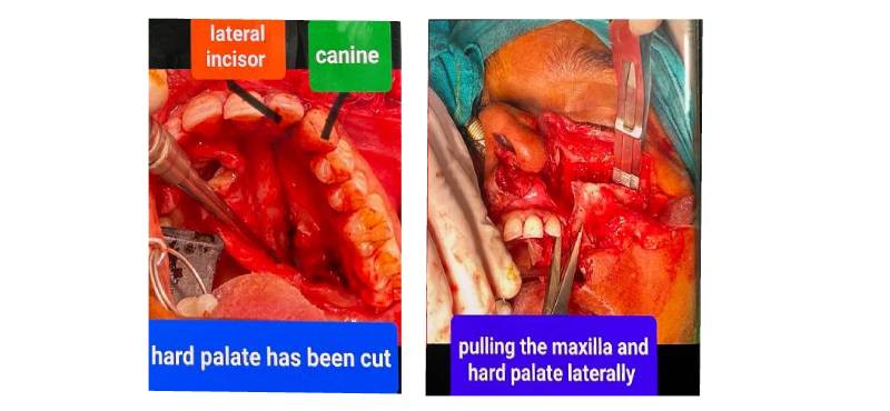
- Swing Maxillotomy: - The second step involved a crucial manoeuvre known as swing maxillotomy. In this intricate process, the left maxilla and hard palate were carefully cut and then swung laterally and upward to expose the tumour. Swing maxillotomy is a specialised surgical technique to enhance access to challenging anatomical sites, such as the pterygopalatine fossa, by temporarily moving the upper jaw away from the skull.
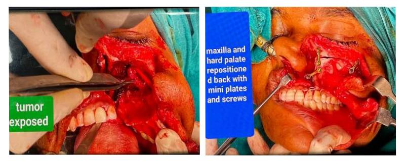
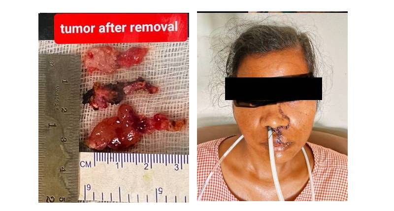
- Gross Total Excision: - Subsequent to tumour exposure, the third step entailed the gross total excision of the tumour. This critical procedure involves completely removing the visible tumour and any surrounding tissues that may harbour abnormal cells. Achieving gross total excision is paramount for minimizing the risk of recurrence and optimizing patient outcomes.
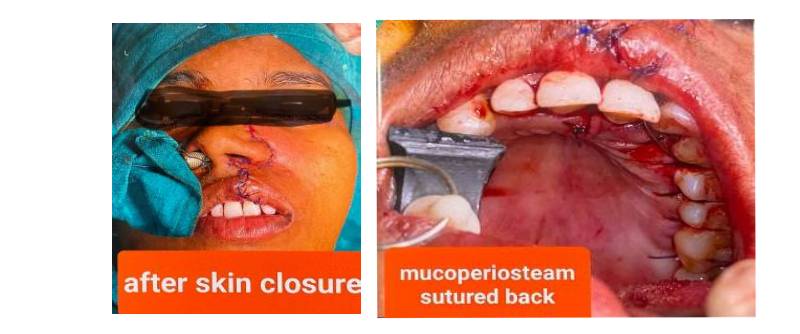
- Repositioning and Stabilization: - Finally, the repositioning of the maxilla and hard palate was executed. These structures were secured back in place using mini plates and screws. This step ensures the restoration of normal facial anatomy and provides structural stability post-surgery.
Meet the Surgeon in Pterygopalatine Fossa Tumour Management: Profiling the Expert Behind Successful Surgeries
Guiding the intricate surgical procedure for the left pterygopalatine fossa tumour was Dr. Mamindla Ravi Kumar, a seasoned Senior Consultant Neurosurgeon specialising in brain and spine surgery. With an impressive academic background encompassing an MBBS, MS, and M.Ch from NIMS, Dr. Kumar's expertise is further augmented by fellowships in Endospine from France and UBE spine surgery.
Additionally, his proficiency in Skull Base Surgery, acquired through a fellowship at MS Ramaiah and WSBF, underscores a commitment to continuous learning and staying abreast of cutting-edge techniques. Dr. Kumar's leadership in this case not only reflects his comprehensive skill set but also his dedication to providing optimal patient care in the intricate realm of neurosurgery.
Conclusion: Achieving Success in Pterygopalatine Fossa Tumour Management
In navigating the complexities of neurosurgery, particularly in addressing rare cases like the pterygopalatine fossa tumour, a delicate blend of expertise, precision, and innovative techniques is paramount. Premier Hospital, Hyderabad stands as a beacon of excellence, emphasizing tailored approaches such as the swing maxillotomy technique. The commitment to optimal patient care is evident in the hospital's team of top-notch medical professionals, making it a multi-speciality hospital of choice in Hyderabad.
All Blogs Categories